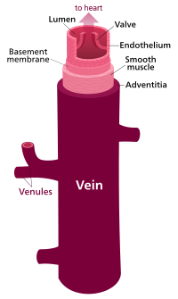
Photo from wikipedia
We read with interest the paper by Zhang et al. investigating the safety and patency of iliac vein stents extending into the inferior vena cava (IVC), which demonstrated a very… Click to show full abstract
We read with interest the paper by Zhang et al. investigating the safety and patency of iliac vein stents extending into the inferior vena cava (IVC), which demonstrated a very low incidence of thrombosis in the contralateral iliac vein (0.55% or 1/183 patients) [1]. The incidence of contralateral deep vein thrombosis (DVT) in this study is remarkably lower than previously published data (1–9.7%) [2, 3], especially as the cohort contained both thrombotic and non-thrombotic iliac vein lesions. Concerns regarding contralateral DVT after common iliac vein (CIV) stent placement in patients with May-Thurner Syndrome (MTS) was recently highlighted by Le et al. [4]. The authors showed that the incidence of contralateral DVT after CIV stent placement was relatively high in their retrospective analysed series [10/111 (9%) patients] and typically occurred late during follow-up (median time of approximately 40 months). They identified that over extension of the CIV stent into the IVC is the main factor determining the development of contralateral DVT and the majority of cases (8/10) were those stents that had extended into the IVC more than 10 mm beyond the iliac confluence and contacting the contralateral wall of the IVC. Apart from causing the “jailing” phenomenon, whereby contralateral venous flow is disturbed, venous intimal hyperplasia has also been proposed as a more likely explanation of contralateral backflow venous obstruction, triggered by persistent irritation of the IVC wall by the overextended stent struts [5]. Although the incidence of contralateral DVT in this study is in keeping with previously published data (1–9.7%), the population of patients studied is more homogeneous than the other referenced series in that only nonthrombotic iliac lesions with MTS were examined. These are the type of lesions that tend to do better with stenting with longer term stent patency demonstrated over post-thrombotic syndrome cases [6] without the need for anti-coagulation therapy. Even with non-thrombotic lesions, this study shows the real danger of caging the contralateral side with a stent over-extended into the IVC. However, as highlighted by the authors, there is still a lack of expert consensus on the ideal location to place a stent in the case of a stenosis adjacent to the iliac confluence. This makes the current study findings of Zhang et al. even more astounding [1]. We appreciate that if the stenotic lesion is not covered sufficiently the stent potentially may migrate caudally or be compressed into a tapering cone (“watermelon seeding”), such that restenosis occurs relatively early post stent deployment. Conversely, there are advocates who propose that the stent is overextended deliberately across the iliac confluence to minimise these concerns with no safety concerns. Part of the controversy regarding the ideal stent location has been the lack of dedicated venous stents to address this problem until recently. We note that that WallstentTM (Boston Scientific, Natick, MA, US) and SMART nitinol stents (Cordis, Bridgewater, NJ, US) were the stents used in this series. The WallstentTM is now relatively old and from personal experience of the senior authors (TYT and YKT) tends to fore-shorten after deployment and post-dilatation and it is * T. Y. Tang [email protected]
Journal Title: Journal of Thrombosis and Thrombolysis
Year Published: 2019
Link to full text (if available)
Share on Social Media: Sign Up to like & get
recommendations!