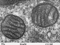
Photo from wikipedia
PurposeTo develop a tool to measure the pH at the surfaces of individual cells.ProceduresThe SNARF pH-sensitive dye was conjugated to a pHLIP® peptide (pH-Low Insertion Peptide) that binds cellular membranes… Click to show full abstract
PurposeTo develop a tool to measure the pH at the surfaces of individual cells.ProceduresThe SNARF pH-sensitive dye was conjugated to a pHLIP® peptide (pH-Low Insertion Peptide) that binds cellular membranes in tumor spheroids. A beam splitter allows simultaneous recording of two images (580 and 640 nm) by a CCD camera. The ratio of the two images is converted into a pH map resolving single spheroid cells. An average pH for each cell is calculated and a pH histogram is derived.ResultsSurface pH depends on cellular glycolytic activity, which was varied by adding glucose or deoxy-glucose. Glucose was found to decrease the surface pH relative to the pH of the bulk solution. The surface pH of metastatic cancer cells was lower than that of non-metastatic cells indicating a higher glycolytic activity.ConclusionsOur method allows cell surface pH measurement and its correlation with cellular glycolytic activity.
Journal Title: Molecular Imaging and Biology
Year Published: 2019
Link to full text (if available)
Share on Social Media: Sign Up to like & get
recommendations!