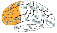
Photo from wikipedia
The prototype disease of Cu toxicity in human is Wilson disease, and cognitive impairment is the presenting symptom of it. There is no study correlating Cu-induced excitotoxicity, apoptosis, and astrocytic… Click to show full abstract
The prototype disease of Cu toxicity in human is Wilson disease, and cognitive impairment is the presenting symptom of it. There is no study correlating Cu-induced excitotoxicity, apoptosis, and astrocytic reaction with memory dysfunction. We report excitotoxicity, apoptosis, and astrocytic reaction of the hippocampus and frontal cortex with memory dysfunction in rat model of Cu toxicity. Thirty-six rats were divided into group I (control) and group II (100 mg/kgBwt/day CuSO4 orally). Y-maze was performed for memory and learning at 0, 30, 60, and 90 days. Frontal and hippocampal free Cu concentration, oxidative stress markers [glutathione (GSH), total antioxidant toxicity (TAC), and malondialdehyde (MDA)], and glutamate were measured by atomic absorption spectroscopy, spectrophotometry, and ELISA, respectively. N-methyl-d-aspartate receptors (NMDARs) NR1, NR2A, and NR2B were done by real-time polymerase chain reaction. Immunohistochemistry for caspase-3 and glial fibrillary acidic protein (GFAP) were done and quantified using the ImageJ software. The glutamate level in hippocampus was increased, and NMDAR expression was decreased at 30, 60, and 90 days in group II compared to group I. In the frontal cortex, glutamate was increased at 90 days, but NMDARs were not significantly different in group II compared to group I. Caspase-3 and GFAP expressions were also higher in group II compared to group I, and these changes were more marked in hippocampus than frontal cortex. These changes correlated with respective free tissue Cu, oxidative stress, and Y-maze attention score. Cu toxicity induces apoptosis and astrocytosis of the hippocampus and frontal cortex through direct or glutamate and oxidative stress pathways, and results in impaired memory and learning.
Journal Title: Molecular Neurobiology
Year Published: 2017
Link to full text (if available)
Share on Social Media: Sign Up to like & get
recommendations!