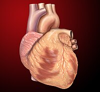
Photo from wikipedia
Cardiac amyloidosis is characterized by the abnormal deposition of misfolded proteins in the heart. A characteristic feature of amyloid proteins are the wrinkled ß-pleated sheets formed within infiltrated tissues. In… Click to show full abstract
Cardiac amyloidosis is characterized by the abnormal deposition of misfolded proteins in the heart. A characteristic feature of amyloid proteins are the wrinkled ß-pleated sheets formed within infiltrated tissues. In the majority of cases, cardiac amyloid can be attributed to light chain amyloid (AL) or transthyretin amyloid (ATTR) protein accumulation. ATTR may be further sub-classified as wild type (senile) or gene mutation (familial). Clinically, ATTR and AL can be similar and distinguishing between them may be a challenge. Scintigraphy using technetium-99m (Tc) labeled bone tracers is helpful in detecting cardiac amyloid and may help differentiate between ATTR and AL cardiac amyloid. ATTR cardiac amyloid typically demonstrates avid myocardial uptake of Tc-labeled bone tracers, whereas in AL cardiac amyloid, cardiac uptake if present, is characteristically low. On occasion however a cardiac amyloid SPECT study using a Tc-labeled bone tracer may be equivocal for ATTR amyloid as the low myocardial uptake of tracer could represent either ATTR or AL amyloid. Current American Society of Nuclear Cardiology practice points for Tc-pyrophosphate imaging suggest image acquisition at 1 hour after tracer injection and at 3 hours if significant blood pool is seen on the 1 hour images. Images are reviewed using a visual grading approach proposed by Perugini et al and a semi-quantitative heart to contra-lateral lung (H/CL) ratio (Figure 1). ATTR amyloid is strongly suggested by a Perugini grade of[ 1 and a H/CL ratio[ 1.5. Equivocal studies have H/CL ratios of 1-1.5 and/or Perugini grade of 0-1.
Journal Title: Journal of Nuclear Cardiology
Year Published: 2019
Link to full text (if available)
Share on Social Media: Sign Up to like & get
recommendations!