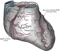
Photo from wikipedia
A 55-year-old woman presented with chest pain with irradiating pain to the left shoulder, nausea, and fatigue for three days. She presented to the emergency department of our hospital. Electrocardiogram… Click to show full abstract
A 55-year-old woman presented with chest pain with irradiating pain to the left shoulder, nausea, and fatigue for three days. She presented to the emergency department of our hospital. Electrocardiogram showed a left bundle branch block and serum troponin T level was slightly elevated (Figure 1). We performed emergency coronary angiography (CAG), which revealed severe stenosis with thrombolysis in myocardial infarction (TIMI) grade 2 flow in the first major septal branch of the left anterior descending artery (LAD) (Figure 2). We started taking her the antiplatelet therapy. Contrastenhanced computed tomography (CT) showed hypoenhancement areas in the basal septal myocardium on the next day (Figure 3a, b). Dual-isotope single-photon emission computed tomography (SPECT) with Tcpyrophosphate (99m-PYP)/TI-chloride showed that the basal to mid-septal wall had high uptake of 99mPYP and perfusion defects on Tl-201 imaging, which corresponded to the blood vessel region of the septal branch (Figures 4, 5). DISCUSSION
Journal Title: Journal of Nuclear Cardiology
Year Published: 2020
Link to full text (if available)
Share on Social Media: Sign Up to like & get
recommendations!