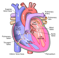
Photo from wikipedia
A 62-year-old female with a known history of pulmonary disease (congenital bronchiectasis of the left lung) presented with right-sided heart failure. Echocardiography demonstrated straightened, restricted tricuspid leaflets fixed in an… Click to show full abstract
A 62-year-old female with a known history of pulmonary disease (congenital bronchiectasis of the left lung) presented with right-sided heart failure. Echocardiography demonstrated straightened, restricted tricuspid leaflets fixed in an almost opened position (Fig. 1a), resulting in severe tricuspid regurgitation (Fig. 1b, Movie Clip 1). Although the normal concave curvature of the tricuspid leaflets was decreased, the tricuspid leaflets and tendons appeared nonthickened on visual assessment. The pulmonic valve was intact. The right heart chambers were moderately dilated and right ventricular contractility was preserved. Pulmonary artery systolic pressure could not be reliably estimated due to lack of coaptation of tricuspid leaflets. These findings were more in favor of organic tricuspid valve disease than cor pulmonale, and hence the suspicion for carcinoid heart disease was raised. Abdominal CT scan revealed a mass (presumably a neuroendocrine tumor) in the right ovary, ascites and abdominal lymphadenopathy. No focal liver lesions were detected. Serum serotonin level was significantly increased to >1000 ng/mL (normal range 80–400 ng/mL). The patient received therapy for right-sided heart failure which resulted in symptomatic improvement, and subsequently was referred to surgery for tricuspid valve replacement. Neuroendocrine tumors (NET), formerly termed “carcinoids”, cause carcinoid heart disease in approximately 25% of cases [1]. Carcinoid heart disease typically develops in the presence of liver metastases as the latter allow serotonin released by the NET cells to reach the heart without being inactivated by monoamine oxidases in the liver and lungs [2]. In the present case, carcinoid heart disease developed early in the absence of liver metastases because of ovarian location of the NET; this permitted vasoactive substances to enter into the right atrium through the ovarian vein and inferior vena cava, bypassing the portal venous system [3]. Although this case is limited by lack of histopathologic findings of the excised valves and the unknown outcome of the patient following surgery, it re-emphasizes the role of echocardiography in raising suspicion for NET [4]; despite co-existing congenital pulmonary disease which potentially could result in right-sided heart failure, typical tricuspid valve morphology with leaflets fixed in an opened position resulting in severe tricuspid regurgitation was pathognomonic of carcinoid heart disease. We also assume that straightened and restricted but visually non-thickened tricuspid leaflets, as well as the intact pulmonic valve, represent an early stage of carcinoid heart disease in this patient.
Journal Title: Journal of Echocardiography
Year Published: 2017
Link to full text (if available)
Share on Social Media: Sign Up to like & get
recommendations!