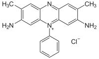
Photo from wikipedia
We report herein a new dual-color fluorescent aptasensor for detection of tumor marker mucin 1 (MUC1) and targeted imaging of MCF-7 cancer cells based on the specific interaction between MUC1… Click to show full abstract
We report herein a new dual-color fluorescent aptasensor for detection of tumor marker mucin 1 (MUC1) and targeted imaging of MCF-7 cancer cells based on the specific interaction between MUC1 and its aptamer S2.2. The aptasensor was prepared by covalent attachment of the cyanine (Cy5)-tagged aptamer S2.2 to fluorescent silicon nanodot (SiND). The fluorescence of S2.2-Cy5 could be quenched by the SiND carrier in the absence of MUC1, and its fluorescence was restored in the presence of MUC1 due to structure switching of S2.2. This aptasensor exhibits specificity for MUC1-possitive MCF-7 cells rather than MUC1-negative MCF-10A cells and Vero cells. The SiND plays multiple roles in this fluorescence assay, making the method easier compared with other approaches. The limit of detection and precision of this method for MUC1 was 1.52 nM and 3.6% (10 nM, n = 7), respectively. The linear range was 3.33-250 nM, and the recoveries in spiked human serum were in the range of 87-108%. This is a simple, selective, sensitive and reliable method, which can well achieve not only quantitative analysis of tumor marker but also dual-color visualization of single cancer cells.
Journal Title: Analytica chimica acta
Year Published: 2018
Link to full text (if available)
Share on Social Media: Sign Up to like & get
recommendations!