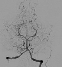
Photo from wikipedia
A 22-year-old male was referred for ocular hypertension workup On presentation his visual acuity was 20/20 in both eyes with normal slit lamp exam. His dilated fundus exam revealed normal… Click to show full abstract
A 22-year-old male was referred for ocular hypertension workup On presentation his visual acuity was 20/20 in both eyes with normal slit lamp exam. His dilated fundus exam revealed normal findings in the right eye. The left eye exhibited retinal vascular changes with telangiectasias and a small amount of surrounding exudation in the superior periphery. An optical coherence tomogram (OCT) through the lesion revealed vascular lumens within the retina (Fig. 1A). Fundus photography revealed an anomalous course of a sclerotic inferior arcade vessel (Fig. 1B). Fluorescein angiography highlighted the course of this vessel (Fig. 1C) with late leakage of the superior vascular region (Fig. 1D). The lesion was observed, but after the exudates were found to be increasing, the patient was treated with laser photocoagulation to the area of leakage. At 2 year follow up the lesion was stable.
Journal Title: American Journal of Ophthalmology Case Reports
Year Published: 2022
Link to full text (if available)
Share on Social Media: Sign Up to like & get
recommendations!