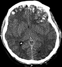
Photo from wikipedia
Abstract Objective This systematic review aimed to synthesize early data on typology and topography of brain abnormalities in adults with COVID-19 in acute/subacute phase. Methods We performed systematic literature search… Click to show full abstract
Abstract Objective This systematic review aimed to synthesize early data on typology and topography of brain abnormalities in adults with COVID-19 in acute/subacute phase. Methods We performed systematic literature search via PubMed, Google Scholar and ScienceDirect on articles published between January 1 and July 05, 2020, using the following strategy and key words: ((covid[Title/Abstract]) OR (sars-cov-2[Title/Abstract]) OR (coronavirus[Title/Abstract])) AND (brain[Title/Abstract]). A total of 286 non-duplicate matches were screened for original contributions reporting brain imaging data related to SARS-Cov-2 presentation in adults. Results The selection criteria were met by 26 articles (including 21 case reports, and 5 cohort studies). The data analysis in a total of 361 patients revealed that brain abnormalities were noted in 124/361 (34%) reviewed cases. Neurologic symptoms were the primary reason for referral for neuroimaging across the studies. Modalities included CT (-angiogram, -perfusion, -venogram), EEG, MRI (-angiogram, functional), and PET. The most frequently reported brain abnormalities were brain white matter (WM) hyperintensities on MRI 66/124 (53% affected cases) and hypodensities on CT (additional 23% affected cases), followed by microhemorrhages, hemorrhages and infarcts, while other types were found in <5% affected cases. WM abnormalities were most frequently noted in bilateral anterior and posterior cerebral WM (50% affected cases). Conclusion About a third of acute/subacute COVID-19 patients referred for neuroimaging show brain abnormalities suggestive of COVID-19-related etiology. The predominant neuroimaging features were diffuse cerebral WM hypodensities / hyperintensities attributable to leukoencephalopathy, leukoaraiosis or rarefield WM.
Journal Title: Brain, Behavior, and Immunity
Year Published: 2020
Link to full text (if available)
Share on Social Media: Sign Up to like & get
recommendations!