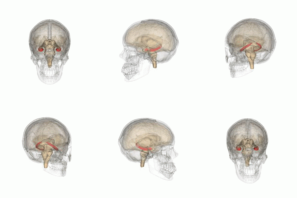
Photo from wikipedia
Neonatal brain injury from hypoxia-ischemia (HI) causes major morbidity. Piglet HI is an established method for testing neuroprotective treatments in large, gyrencephalic brain. Though many neurobehavior tests exist for rodents,… Click to show full abstract
Neonatal brain injury from hypoxia-ischemia (HI) causes major morbidity. Piglet HI is an established method for testing neuroprotective treatments in large, gyrencephalic brain. Though many neurobehavior tests exist for rodents, such tests and their associations with neuropathologic injury remain underdeveloped and underutilized in large, neonatal HI animal models. We examined whether spatial T-maze and inclined beam tests distinguish cognitive and motor differences between HI and sham piglets and correlate with neuropathologic injury. Neonatal piglets were randomized to whole-body HI or sham procedure, and they began T-maze and inclined beam testing 17 days later. HI piglets had more incorrect T-maze turns than did shams. Beam walking time did not differ between groups. Neuropathologic evaluations at 33 days validated the injury with putamen neuron loss after HI to below that of sham procedure. HI decreased the numbers of CA3 pyramidal neurons but not CA1 pyramidal neurons or dentate gyrus granule neurons. Though the number of hippocampal parvalbumin-positive interneurons did not differ between groups, HI reduced the number of CA1 interneuron dendrites. Piglets with more incorrect turns had greater CA3 neuron loss, and piglets that took longer in the maze had fewer CA3 interneurons. The number of putamen neurons was unrelated to T-maze or beam performance. We conclude that neonatal HI causes hippocampal CA3 neuron loss, CA1 interneuron dendritic attrition, and putamen neuron loss at 1-month recovery. The spatial T-maze identifies learning and memory deficits that are related to loss of CA3 pyramidal neurons and fewer parvalbumin-positive interneurons independent of putamen injury.
Journal Title: Behavioural Brain Research
Year Published: 2019
Link to full text (if available)
Share on Social Media: Sign Up to like & get
recommendations!