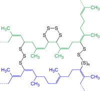
Photo from wikipedia
The T cell antigen receptor (TCR) binds a ligand consisting of antigenic peptide bound to a cell surface molecule encoded by the major histocompatibility complex (pMHC) to begin the process… Click to show full abstract
The T cell antigen receptor (TCR) binds a ligand consisting of antigenic peptide bound to a cell surface molecule encoded by the major histocompatibility complex (pMHC) to begin the process of T cell activation. TCR engagement leads to protein tyrosine kinase (PTK) recruitment to the TCR and PTK activation, and activated PTKs phosphorylate the membrane-bound adapter protein LAT and the cytosolic adapter protein SLP-76. Phosphorylated LAT and SLP-76 recruit a number of other adapters and enzymes including GADS, PLCγ, GRB2, NCK, VAV and ITK. The protein complexes nucleating at LAT are highly heterogeneous, and the formation of these complexes is highly co-operative. Different LAT complexes activate different effector function. For example, the LAT/GADS/SLP-76 complex recruits the PTK ITK to activate PLCγ, leading to Ca2+ flux and MAPK activation. Here we study the LAT-GADS-SLP-76 tri-molecular complex by generating it in vitro from purified component proteins and a LAT phospho-peptide. Isolation of this complex and others should enable generation of structural information, which would be required to design specific inhibitors for modulation of signaling.
Journal Title: Biophysical Journal
Year Published: 2017
Link to full text (if available)
Share on Social Media: Sign Up to like & get
recommendations!