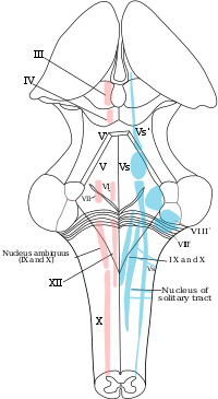
Photo from wikipedia
The pain sensation system is highly conserved among species, thus animal models have been used to investigate relevant tissues. The focus for head-specific pain has been on the primary nociceptive… Click to show full abstract
The pain sensation system is highly conserved among species, thus animal models have been used to investigate relevant tissues. The focus for head-specific pain has been on the primary nociceptive neurons in the trigeminal pathway, i.e. trigeminal ganglia. The secondary nociceptive neurons of the trigeminal pathway, trigeminal nucleus caudalis (TNC), have not been assessed. We expect different gene expression profiles compared to the homologous spinal cord dorsal horn (SDH), as several signalling substances provoke head-specific pain but not peripheral pain. We aim to provide expression profiles of TNC and SDH, tissues highly relevant for pain- and migraine-studies. We extracted RNA from laser capture microdissected laminae I-V from TNC and SDH from six Wistar rats for RNA-Sequencing. We showed the expression profiles of genes involved in neural signal transmission and found that among all G protein-coupled receptors Gabbr1 was highest expressed in both tissues. Among the migraine-associated genes we showed that Cacna1a, where non-synonyms mutations can cause familial hemiplegic migraine, was highly expressed with a slightly lower expression in TNC than in SDH. To show the genetic differences between the two homologous systems we performed a differential expression analysis, revealing 1696 genes higher and 1895 genes lower expressed genes in TNC than in SDH, of which many were neuronal-related. The high number of differentially expressed genes shows the large genetic difference between the trigeminal and spinothalamic system. Our results contribute to the characterization of nociceptive pathways, which may help us understanding why several signalling molecules cause headache and no peripheral pain.
Journal Title: Brain Research
Year Published: 2018
Link to full text (if available)
Share on Social Media: Sign Up to like & get
recommendations!