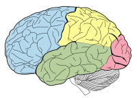
Photo from wikipedia
PURPOSE To evaluate temporal changes in gamma-aminobutyric acid (GABA) signals in the hippocampus during epileptiform activity induced by kainic acid (KA) in a rat model of status epilepticus using chemical… Click to show full abstract
PURPOSE To evaluate temporal changes in gamma-aminobutyric acid (GABA) signals in the hippocampus during epileptiform activity induced by kainic acid (KA) in a rat model of status epilepticus using chemical exchange saturation transfer (CEST) imaging technique. METHODS CEST imaging and 1H magnetic resonance spectroscopy (1H MRS) were applied to a systemic KA-induced rat model to compare GABA signals. All data acquisition and analytical procedures were performed at three different time points (before KA injection, and 1 and 3 h after injection). The CEST signal was analyzed based on regions of interests (ROIs) in the hippocampus, while 1H MRS was analyzed within a 12.0 μL ROI in the left hippocampus. Signal correlations between the two methods were evaluated as a function of time change up to 3 h after KA injection. RESULTS The measured GABA CEST-weighted signal intensities of the rat epileptic hippocampus before injection showed significant differences from those after (averaged signals from both hippocampi: 4.37% ± 0.87% and 7.305 ± 1.11%; P < 0.05), although the signal had increased slightly at both time points after KA injection, the differences were not significant (P > 0.05). In contrast, the correlation between the CEST imaging values and 1H MRS was significant (r ≥ 0.64; P < 0.05; in all cases). CONCLUSIONS GABA signal changes during epileptiform activity in the rat hippocampus, as detected using CEST imaging, provided a significant contrast according to changes in metabolic activity. Our technical approach may serve as a potential supplemental option to provide biomarkers for brain disease.
Journal Title: Brain Research
Year Published: 2019
Link to full text (if available)
Share on Social Media: Sign Up to like & get
recommendations!