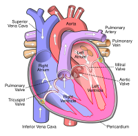
Photo from wikipedia
Background Right atrial pressure (RAP) and intravascular volume status are estimated by echocardiography with 2D imaging of inferior vena cava (IVC) diameter and collapsibility. 3D imaging can avoid misalignment and… Click to show full abstract
Background Right atrial pressure (RAP) and intravascular volume status are estimated by echocardiography with 2D imaging of inferior vena cava (IVC) diameter and collapsibility. 3D imaging can avoid misalignment and likely achieve more accurate length measurements. We sought to compare the diagnostic performance of 2D- and 3D-derived IVC measurements with regard to clinical parameters associated with heart failure (HF). We also assessed the degree of discordance between the two methods. Methods 200 patients had 2D and 3D subcostal IVC imaging performed before and after inspiratory sniff. Images were acquired using Philips iE33 and EPIQ7 systems and analyzed using QLAB v10.3 (Andover, MA). RAP was categorized as normal, mildly elevated, or elevated according to 2015 ASE guidelines. The reference 2D diameter limit was 2.1 cm and corresponding 3D-derived cross-sectional major axis (2.7 cm) and minor axis (1.9 cm) limits were obtained by regression analysis. Collapsibility index (CI) was calculated along each 2D and 3D axis direction. Sphericity index (SI) was calculated as the ratio of minor to major axis lengths and IVCs were categorized as either spherical (SI ≥ 0.7) or elliptical (SI Results 2D IVC diameter was 2.0 ± 0.6 cm, while 3D major and minor axes were 2.7 ± 0.7 cm and 1.9 ± 0.6 cm. 2D RAP category concordance with 3D major and minor axis RAP categories was 43% and 63%, respectively. CI was different along 3D major (44 ± 19%) and minor axes (52 ± 21%; p Table 1 . Conclusions Echocardiographic 2D- and 3D-derived categorization of RAP was discordant in roughly half of the analyzed cases. Collapsibility differs along the major and minor axes of the IVC. Vessel sphericity influences the discrepancies between 2D and 3D measurements. Furthermore, 3D cross-sectional IVC area outperformed standard 2D IVC diameter for predicting certain clinical variables. Since the IVC is typically dynamic and often non-cylindrical in shape, cross-sectional measurements by 3D imaging may provide greater fidelity in classifying RAP.
Journal Title: Journal of Cardiac Failure
Year Published: 2018
Link to full text (if available)
Share on Social Media: Sign Up to like & get
recommendations!