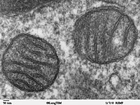
Photo from wikipedia
SUMMARY Genetic and biochemical defects of mitochondrial function are a major cause of human disease, but their link to mitochondrial morphology in situ has not been defined. Here, we develop… Click to show full abstract
SUMMARY Genetic and biochemical defects of mitochondrial function are a major cause of human disease, but their link to mitochondrial morphology in situ has not been defined. Here, we develop a quantitative three-dimensional approach to map mitochondrial network organization in human muscle at electron microscopy resolution. We establish morphological differences between human and mouse and among patients with mitochondrial DNA (mtDNA) diseases compared to healthy controls. We also define the ultrastructure and prevalence of mitochondrial nanotunnels, which exist as either free-ended or connecting membrane protrusions across non-adjacent mitochondria. A multivariate model integrating mitochondrial volume, morphological complexity, and branching anisotropy computed across individual mitochondria and mitochondrial populations identifies increased proportion of simple mitochondria and nanotunnels as a discriminant signature of mitochondrial stress. Overall, these data define the nature of the mitochondrial network in human muscle, quantify human-mouse differences, and suggest potential morphological markers of mitochondrial dysfunction in human tissues.
Journal Title: Cell reports
Year Published: 2019
Link to full text (if available)
Share on Social Media: Sign Up to like & get
recommendations!