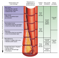
Photo from wikipedia
We evaluated the virtual monochromatic imaging (VMI) energy levels that maximize image quality of each coronary plaque component in dual-energy computed tomography angiography in 495 coronary segments (45 for each… Click to show full abstract
We evaluated the virtual monochromatic imaging (VMI) energy levels that maximize image quality of each coronary plaque component in dual-energy computed tomography angiography in 495 coronary segments (45 for each energy level). Maximal signal-to-noise ratios were different for plaque, lumen, fat, and surrounding tissue (p<0.05). Maximal contrast-to-noise ratios were observed at 70keV for calcified plaque (CP), non-calcified plaque (NCP), and fat in comparison with the lumen (p<0.05), and 70keV and 120keV for NCP in comparison with fat (p=0.144). VMI demonstrated maximal image quality at different energy levels for each component of coronary artery plaque.
Journal Title: Clinical imaging
Year Published: 2017
Link to full text (if available)
Share on Social Media: Sign Up to like & get
recommendations!