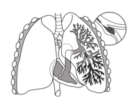
Photo from wikipedia
PURPOSE Limited diagnostic options exist for patients with suspected pulmonary embolism (PE) who cannot undergo CT-angiogram (CTA). CT-ventilation methods recover respiratory motion-induced lung volume changes as a surrogate for ventilation.… Click to show full abstract
PURPOSE Limited diagnostic options exist for patients with suspected pulmonary embolism (PE) who cannot undergo CT-angiogram (CTA). CT-ventilation methods recover respiratory motion-induced lung volume changes as a surrogate for ventilation. We recently demonstrated that pulmonary blood mass change, induced by tidal respiratory motion, is a potential surrogate for pulmonary perfusion. In this study, we examine blood mass and volume change in patients with PE and parenchymal lung abnormalities (PLA). METHODS A cross-sectional analysis was conducted on a prospective, cohort-study with 129 consecutive PE suspected patients. Patients received 4DCT within 48 h of CTA and were classified as having PLA and/or PE. Global volume change (VC) and percent global pulmonary blood mass change (PBM) were calculated for each patient. Associations with disease type were evaluated using quantile regression. RESULTS 68 of 129 patients were PE positive on CTA. Median change in PBM for PE-positive patients (0.056; 95% CI: 0.045, 0.068; IQR: 0.051) was smaller than that of PE-negative patients (0.077; 95% CI: 0.064, 0.089; IQR: 0.056), with an estimated difference of 0.021 (95% CI: 0.003, 0.038; p = 0.0190). PLA was detected in 57 (44.2%) patients. Median VC for PLA-positive patients (1.26; 95% CI: 1.22, 1.30; IQR: 0.15) showed no significant difference from PLA-negative VC (1.25; 95% CI: 1.21, 1.28; IQR: 0.15). CONCLUSIONS We demonstrate that pulmonary blood mass change is significantly lower in PE-positive patients compared to PE-negative patients, indicating that PBM derived from dynamic non-contrast CT is a potentially useful surrogate for pulmonary perfusion.
Journal Title: Clinical imaging
Year Published: 2021
Link to full text (if available)
Share on Social Media: Sign Up to like & get
recommendations!