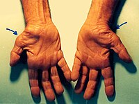
Photo from wikipedia
Introduction Ulnar nerve is frequently involved nerve in the upper limb producing mononeuropathy and on many occasions it is not explored beyond electrophysiology. The aim of the study here is… Click to show full abstract
Introduction Ulnar nerve is frequently involved nerve in the upper limb producing mononeuropathy and on many occasions it is not explored beyond electrophysiology. The aim of the study here is to present the electrophysiology and the magnetic resonance imaging (MRI) findings of non-traumatic ulnar neuropathies. Methods In this prospective study 21 consecutive patients presenting with non-traumatic ulnar neuropathy were examined clinically. Nerve conductions were performed in all the four limbs while EMG was done in the muscles supplied by the involved ulnar nerve and other muscles only if required. This was followed by MRI of the suspected site of lesion in the course of the nerve. Results All the patients had clinical findings suggestive of ulnar neuropathy. The NCV-EMG localized a lesion in 20 ulnar nerves where as it was normal in 2. MRI revealed a lesion in 19 (86%) of the total 22 ulnar nerves studied. The causative lesions identified using MRI were - Ulnar neuritis (n = 7), enlarged cubital node at elbow (n = 3), Synovial cyst at Guyan’s canal (n = 2), arthritis of ulnohumeral joint (n = 2), # medial epicondyle (n = 1), Accessory muscle in cubital tunnel (n = 1), Koch’s of ulnohumeral joint (n = 1) and Neurofibroma (n = 2). Conclusion Ulnar neuritis is the most common cause of non-traumatic ulnar neuropathy followed by enlarged cubital node and neurofibroma in our series. MRI offers a noninvasive method for identifying an etiology of lesion along the nerve course and is likely to modify the management decision.
Journal Title: Clinical Neurophysiology
Year Published: 2018
Link to full text (if available)
Share on Social Media: Sign Up to like & get
recommendations!