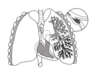
Photo from wikipedia
PURPOSE To compare the quantitative and qualitative lung perfusion data acquired with dual energy CT (DECT) to that acquired with a large field-of-view cadmium-zinc-telluride camera single-photon emission CT coupled to… Click to show full abstract
PURPOSE To compare the quantitative and qualitative lung perfusion data acquired with dual energy CT (DECT) to that acquired with a large field-of-view cadmium-zinc-telluride camera single-photon emission CT coupled to a CT system (SPECT-CT). MATERIALS AND METHODS A total of 53 patients who underwent both dual-layer DECT angiography and perfusion SPECT-CT for pulmonary hypertension or pre-operative lobar resection surgery were retrospectively included. There were 30 men and 23 women with a mean age of 65.4±17.5 (SD)years (range: 18-88years). Relative lobar perfusion was calculated by dividing the amount (of radiotracer or iodinated contrast agent) per lobe by the total amount in both lungs. Linear regression, Bland-Altman analysis, and Pearson's correlation coefficient were also calculated. Kappa test was used to test agreements in morphology and severity of perfusion defects assessed on SPECT-CT and on DECT iodine maps with a one-month interval. Wilcoxon rank sum test was used to compare the sharpness of perfusion defects and radiation dose among modalities. RESULTS Strong correlations for relative lobar perfusion using linear regression analysis and Pearson's correlation coefficient (r=0.93) were found. Bland-Altman analysis revealed a -0.10 bias, with limits of agreement between [-6.01; 5.81]. With respect to SPECT- CT as standard of reference, the sensitivity, specificity, PPV, NPV, accuracy for lobar perfusion defects were 89.4% (95% CI: 82.6-93.4%), 96.5% (95% CI: 92.1-98.5%), 95.6% (95% CI: 90.9-97.8%), 91.4% (95% CI: 85.6-94.9%) and 93.0% (95% CI: 87.6-96.1%) respectively. High level of agreement was found for morphology and severity of perfusion defects between modalities (Kappa=0.84 and 0.86 respectively) and on DECT images among readers (Kappa=0.94 and 0.89 respectively). A significantly sharper delineation of perfusion defects was found on DECT images (P<0.0001) using a significantly lower equivalent dose of 4.1±2.3 (SD) mSv (range: 1.9-11.85mSv) compared to an equivalent dose of 5.3±1.1 (SD) mSv (range: 2.8-7.3mSv) for SPECT-CT, corresponding to a 21.2% dose reduction (P=0.0004). CONCLUSION DECT imaging shows strong quantitative correlations and qualitative agreements with SPECT-CT for the evaluation of lung perfusion.
Journal Title: Diagnostic and interventional imaging
Year Published: 2020
Link to full text (if available)
Share on Social Media: Sign Up to like & get
recommendations!