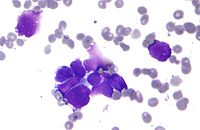
Photo from wikipedia
Overexpression of the Bcl-2 protein has emerged as a hallmark of carcinoma cells and can be employed as a biochemical biomarker of these cells. Therefore, some Bcl-2 protein fluorescence probes… Click to show full abstract
Overexpression of the Bcl-2 protein has emerged as a hallmark of carcinoma cells and can be employed as a biochemical biomarker of these cells. Therefore, some Bcl-2 protein fluorescence probes (BPFPs) were designed for Bcl-2 protein quantification and carcinoma cells labeling. The high Bcl-2 protein binding affinity (Ki < 1 nM) and selectivity (over 50,000-fold Bcl-2 protein selectivity against Mcl-1 protein) of BPFP1 endow it with the ability to detect trace amounts of Bcl-2 protein. After being incubated with a range of concentrations of Bcl-2 protein, BPFP1 exhibited the desired fluorescence properties and its fluorescence intensity is proportional to Bcl-2 protein concentration. Therefore, BPFP1 provides a convenient approach for Bcl-2 protein quantification and we could determine the concentration of Bcl-2 protein based on the BPFP1's fluorescence intensity. Subsequent studies revealed that BPFP1 can fluorescently label carcinoma cells by binding to overexpressed Bcl-2 protein in living cells, and can distinguish carcinoma cells (HL-60 cells and ACHN cells) from normal-tissue cells (HUVECs) according to the different Bcl-2 protein expression levels between carcinoma cells and normal tissue cells. In the present study, BPFP1 represents a new tool for Bcl-2 protein quantification, carcinoma cell visualization and cell sorting. Moreover, BPFP1 can be used in the future for early cancer diagnosis by detecting carcinoma cells in patient tissues.
Journal Title: European journal of medicinal chemistry
Year Published: 2021
Link to full text (if available)
Share on Social Media: Sign Up to like & get
recommendations!