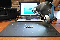
Photo from wikipedia
Purpose Three-dimensional (3D) imaging devices are emerging in operating theatres for intra-operative applications. Although there are reported benefits of this technology, such as increased surgical accuracy and higher patient safety,… Click to show full abstract
Purpose Three-dimensional (3D) imaging devices are emerging in operating theatres for intra-operative applications. Although there are reported benefits of this technology, such as increased surgical accuracy and higher patient safety, radiation exposure of staff and patient remains a concern. This study aims at evaluating this exposure when using the O-arm® (2D and 3D imaging device). Methods To assess patient radiation exposure, absorbed doses to organs were measured on two anthropomorphic phantoms representing a five-year-old child and an adult female. The phantoms are made of slabs containing holes at the location of different organs, where thermoluminescent detectors (TLDs) were placed. Organ doses were measured for one 3D-acquisition of the O-arm® and during one minute of fluoroscopy using a conventional C-arm for comparative purposes. Skin entrance dose rates were also evaluated during 2D fluoroscopy by measuring incident kerma rates on the phantom. Staff exposure was evaluated by creating a 2D ambient equivalent dose cartography of the operating theatre for a single 3D O-arm® acquisition. Moreover, whole body and eye lens doses to the O-arm® technician were measured by placing an anthropomorphic phantom equipped with TLDs. Results Phantom organ doses for one 3D pelvic acquisition with the O-arm® ranged from 4.0 μGy to 6.7 mGy for the adult and from 0.9 μGy to 1.7 mGy for the child; on average 4 times higher than doses obtained after one minute of fluoroscopy with the C-Arm. Consistently, kerma rates at the surface of the skin were between 6 and 164 times lower for the C-Arm compared to the O-arm®. Results from the cartography showed that at 2 m from the isocenter, the maximum ambient equivalent dose is 11 μSv per acquisition and 3 μSv at the technician’s position. Dosimeters placed on the technician phantom showed doses below the detection limit (i.e. 0.1 mSv). Conclusions Doses delivered to the patient during a 3D acquisition of the O-Arm® are systematically higher than the corresponding irradiation with the C-arm. The staff, if positioned as far as possible from the isocenter and adequately equipped with protective gear, may remain inside the operative room during an O-arm® 3D acquisition.
Journal Title: Physica Medica
Year Published: 2018
Link to full text (if available)
Share on Social Media: Sign Up to like & get
recommendations!