Photo from wikipedia
OBJECTIVES To determine what computed tomographic (CT) dimensions can predict obstructive lung disease on routine chest CT scans by comparing morphological and densitometric CT findings with pulmonary function test (PFT)… Click to show full abstract
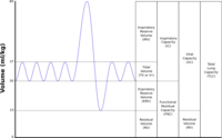
OBJECTIVES To determine what computed tomographic (CT) dimensions can predict obstructive lung disease on routine chest CT scans by comparing morphological and densitometric CT findings with pulmonary function test (PFT) in normal subjects and patients with chronic obstructive pulmonary disease (COPD). MATERIALS AND METHODS Consecutive patients (n = 646; 260 females and 386 males; mean age 54.9 years, ranged 20-90 years) who received chest CT scans with densitometry during a 3-month period were retrospectively analyzed in single center. PFT was undertaken in 235 patients (152 males, 83 females) at same times of CT scanning. The patients were grouped by age (<30 years, 31-45 years, 46-60 years, and >61 years). CT parameters including tracheal, azygoesophageal, thoracic vertical, anterior-posterior (AP), transverse diameters, transverse cardiac diameter, diameters of main, right, and left pulmonary arteries, and CT densitometric values including lung volume and density (-900 to -1000 Hounsfield Units, HU), low attenuation value cluster (default threshold: -950 HU) were compared with PFT values. Spearman correlation coefficients was used to evaluate the relationship between the CT indices and PFT. RESULTS Ninety of 235 patients with PFT were smokers (76 males, 14 females). Obstructive PFT was detected in 65 patients (27.7%: 46 males, 19 females). Male smokers with obstructive PFT displayed significantly larger thoracic anterior-posterior (mean: normal, 172.3 cm versus COPD, 185.9 cm, p = 0.0001) and smaller transverse diameters (mean: normal, 247.0 cm vs. COPD, 235.8 cm, p = 0.01), and increased right pulmonary artery diameter (mean: normal, 20.3 cm v s. COPD, 22.1 cm, p < 0.001), and increased left pulmonary artery diameter (mean: normal, 19.7 cm vs. COPD, 20.6 cm, p < 0.025). The lung parenchyma density (-1000 to -900 HU) and greater concentration of largest cluster on densitometry were significantly different between normal and obstructive PFT pattern in male smoker. Residual volume and total lung capacity are positively correlated with lung volume and lung density (-1000 to -800) of densitometry. CONCLUSIONS CT findings of the overexpansion of the lungs, such as increases in the vertical diameter of the lung and decreases in the transverse diameter of the heart, can be significant as indirect findings of early chronic obstructive diseases. However, despite the significant CT findings in male smokers, particularly those in their 40s, most lung function parameters were not decidedly abnormal.
Journal Title: European journal of radiology
Year Published: 2018
Link to full text (if available)
Share on Social Media: Sign Up to like & get
recommendations!