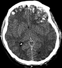
Photo from wikipedia
PURPOSE To test markers from conventional and diffusion Magnetic Resonance Imaging (MRI) as possible predictors of cognitive outcome following rehabilitation therapy in children with acquired brain injury (ABI). METHODS Twenty-one… Click to show full abstract
PURPOSE To test markers from conventional and diffusion Magnetic Resonance Imaging (MRI) as possible predictors of cognitive outcome following rehabilitation therapy in children with acquired brain injury (ABI). METHODS Twenty-one children (10 boys, mean age 11.6 years, range 7.1-19.4) with stroke or traumatic brain injury underwent MRI including Diffusion Tensor Imaging (DTI) before admission to the rehabilitation centre. The conventional images were scored according to a standardised injury scoring system, and mean Fractional Anisotropy (FA) was determined within the Corpus Callosum (CC), as this structure is hypothesised to play an important role in cognition. Both conventional MRI injury scores and mean FA of the CC and its sub-regions were compared with standard functional cognitive outcome scores. Relationships between MRI indices and cognitive outcome scores were assessed using multiple regression and receiver operating characteristic (ROC) analyses. RESULTS A backwards regression analysis revealed that the mean FA of the CC body and genu and the supratentorial injury score appear to represent the best predictors of outcome, together with the age at rehabilitation and time in rehabilitation. In the ROC analysis, the mean FA values of the CC body and genu and the infratentorial injury score provided the highest sensitivity, while the mean FA of the CC splenium showed the highest specificity for outcome. CONCLUSIONS The conventional MRI injury scores and DTI metrics from the CC reflect cognitive outcomes following rehabilitation. Neuroimaging methods such as MRI with DTI may therefore provide important markers for cognitive recovery after brain injury.
Journal Title: European journal of radiology
Year Published: 2020
Link to full text (if available)
Share on Social Media: Sign Up to like & get
recommendations!