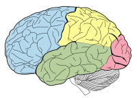
Photo from wikipedia
BACKGROUND Mesial temporal sclerosis (MTS) is the most common cause of temporal lobe epilepsy (TLE). While MTS is associated with a high cure rate after temporal lobectomy (TL), postoperative neurocognitive… Click to show full abstract
BACKGROUND Mesial temporal sclerosis (MTS) is the most common cause of temporal lobe epilepsy (TLE). While MTS is associated with a high cure rate after temporal lobectomy (TL), postoperative neurocognitive deficits are common, and a subset of patients may continue to have refractory seizures. OBJECTIVE To use magnetic resonance (MR) volumetry to identify features of the mesial temporal lobe in patients with MTS that correlate with seizure and neurocognitive outcome after temporal lobectomy. METHODS Thirty-five patients with unilateral MTS, high-resolution MR imaging, and at least one year of postoperative assessments were retrospectively examined. Volumetric analysis of the hippocampus, parahippocampal gyrus (PHG) and FLAIR hyperintensity of the affected temporal lobe was performed. TL resections were manually segmented, and resection heat maps reflecting seizure outcome were produced. The degree of preoperative atrophy of the affected mesial structures relative to the unaffected side were related to preoperative and postoperative component scores of verbal and visuospatial memory as well as confrontation naming. RESULTS Greater FLAIR hyperintense volume was associated with favorable seizure outcome at one year and last follow-up. Resections extending most medial and posteriorly were associated with favorable seizure outcome. In patients with left MTS, less atrophy of the affected PHG was predictive of higher preoperative naming scores and greater postoperative naming deficit, while less hippocampal atrophy was predictive of higher preoperative verbal memory component scores. CONCLUSION Greater hippocampal FLAIR volume is associated with favorable surgical outcome. Hippocampal volume correlates with preoperative verbal memory, while PHG volume is implicated in confrontation naming ability.
Journal Title: Epilepsy Research
Year Published: 2021
Link to full text (if available)
Share on Social Media: Sign Up to like & get
recommendations!