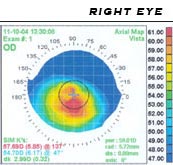
Photo from wikipedia
ABSTRACT Having established a main neuronal origin for noradrenaline (NA) in the cornea, we set out to study the physiologic determinants of its release and to correlate functional findings with… Click to show full abstract
ABSTRACT Having established a main neuronal origin for noradrenaline (NA) in the cornea, we set out to study the physiologic determinants of its release and to correlate functional findings with sympathetic nerve density and overall topography. Whole corneas were obtained from 3 to 4 month‐old rabbits and human donors. Study of prejunctional effects was carried out after incubation with radiolabelled NA (3H‐NA). Corneas were superfused with warm aerated amine‐free medium with cocaine and hydrocortisone to block subsequent neuronal and extraneuronal NA uptake. Samples were collected every 5min. Four periods of transmural electrical stimulation were applied to assess evoked release of 3H‐NA in the absence and in the presence of alpha‐2 adrenoceptor antagonists. Catecholamines were extracted with alumina from the superfusate collected and quantified by high pressure liquid chromatography with electrochemical detection (HPLC‐ED). Corneal nerve morphology was studied by immunofluorescence staining with monoclonal antibodies and subsequent confocal microscopy. Corneal lamellar sections were also produced (epithelium, stroma, endothelium) and endogenous NA and adrenaline (AD) were quantified by HPLC‐ED. Results are means±SEM. ANOVA and t‐tests were used for statistical analysis. Ratios between enzymatic end products and their substrates were calculated. In both rabbit and human corneas, electrical stimulation increased the outflow of 3H‐NA per minute and per shock. Addition of the alpha‐2 adrenoceptor antagonist rauwolscine further increased the electrically‐evoked overflow of 3H‐NA in a concentration‐dependent manner. Immunofluorescence revealed particular staining patterns for sensory and sympathetic fibres, epithelial cells and stromal keratocytes. In human corneal lamellar sections only NA was identified, particularly in the endothelium and epithelium. In the rabbit, concentration of NA was ten times that of AD. Electrically‐evoked overflow reflects action potential‐induced NA release by sympathetic nerves in the cornea and an alpha‐2 adrenoceptor‐mediated mechanism for its release is presented. Sympathetic innervation has similar functional relevance in both rabbit and human corneas. HIGHLIGHTSThe occurrence of an electrically‐evoked overflow of NA reflects action potential‐induced neurotransmitter release by sympathetic nerves.There is alpha‐2 adrenoceptor‐mediated mechanism for neuronal NA release in the cornea.NA predominates significantly over AD in the cornea, particularly in the endothelium (sequestered from aqueous humour) and epithelium (mostly neuronal‐derived, with some uptake by epithelial cells).Sympathetic innervation has similar functional relevance in rabbit and human corneas.Sympathetic fibres are located in the anterior stroma, mostly in the peripheral cornea, and extend to the basal layer of the epithelium; epithelial cells may have hypothetical NA‐synthesising capability.
Journal Title: Experimental Eye Research
Year Published: 2018
Link to full text (if available)
Share on Social Media: Sign Up to like & get
recommendations!