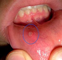
Photo from wikipedia
The aim of this study was to investigate key points for the differential diagnosis of immunoglobulin G4-related sialadenitis (IgG4-RS) and Kimura's disease (KD) involving the salivary glands. The clinical, serological,… Click to show full abstract
The aim of this study was to investigate key points for the differential diagnosis of immunoglobulin G4-related sialadenitis (IgG4-RS) and Kimura's disease (KD) involving the salivary glands. The clinical, serological, radiological, histological, and immunohistochemical features of 85 IgG4-RS cases and 52 KD cases were evaluated comparatively. Seventy-two IgG4-RS cases had enlargement of multiple salivary and/or lacrimal glands; 67 patients had bilateral submandibular gland (SMG) involvement. Unilateral parotid gland involvement (59.6%) and comorbid skin lesions (61.5%) were common in KD. Serum IgG4 was elevated in 94.1% of IgG4-RS cases versus 19.0% of KD cases (cut-off value=266.5mg/dl). KD was more commonly associated with elevated eosinophil counts (86% vs 23.1%) and elevated IgE concentrations (95.5% vs 76.6%). Storiform fibrosis, irregular lymphoid follicles, and increased IgG4-positive cells (112.9±37.6/high-power field (HPF)) were common in IgG4-RS. Acellular fibrosis, regular lymphoid follicles, IgE-positive reticular networks, increased IgE-positive cells (43.4±26.7/HPF), and tryptase-positive mast cells (29.7±13.3/HPF) were usually detected in KD. Computed tomography showed that 85.7% of KD cases involved subcutaneous fat tissue. A superficial hypoechoic and reticular pattern with multiple hypoechoic foci were the sonographic features of the SMG in IgG4-RS. Despite numerous overlapping manifestations, histopathological examination showed meaningful differences in the types of fibrosis, eosinophils, and IgG4-positive cell counts. Comprehensive evaluation of clinical, serological, radiological, and histopathological features are crucial for the differential diagnosis.
Journal Title: International journal of oral and maxillofacial surgery
Year Published: 2020
Link to full text (if available)
Share on Social Media: Sign Up to like & get
recommendations!