Photo from wikipedia
BACKGROUND The diagnosis of penetrating isolated diaphragmatic injuries can be challenging because they are usually asymptomatic. Diagnosis by chest X-ray (CXR) is unreliable, while CT scan is reported to be… Click to show full abstract
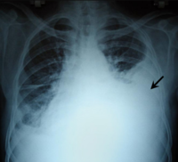
BACKGROUND The diagnosis of penetrating isolated diaphragmatic injuries can be challenging because they are usually asymptomatic. Diagnosis by chest X-ray (CXR) is unreliable, while CT scan is reported to be more valuable. This study evaluated the diagnostic ability of CXR and CT in patients with proven DI. METHODS Single center retrospective study (2009-2019), including all patients with penetrating diaphragmatic injuries (pDI) documented at laparotomy or laparoscopy with preoperative CXR and/or CT evaluation. Imaging findings included hemo/pneumothorax, hemoperitoneum, pneumoperitoneum, elevated diaphragm, definitive DI, diaphragmatic hernia, and associated abdominal injuries. RESULTS 230 patients were included, 62 (27%) of which had isolated pDI, while 168 (73%) had associated abdominal or chest trauma. Of the 221 patients with proven DI and preoperative CXR, the CXR showed hemo/pneumothorax in 99 (45%), elevated diaphragm in 51 (23%), and diaphragmatic hernia in 4 (1.8%). In 86 (39%) patients, the CXR was normal. In 126 patients with pDI and preoperative CT, imaging showed hemo/pneumothorax in 95 (75%), hemoperitoneum in 66 (52%), pneumoperitoneum in 35 (28%), definitive DI in 56 (44%), suspected DI in 26 (21%), and no abnormality in 3 (2%). Of the 57 patients with isolated pDI the CXR showed a hemo/pneumothorax in 24 (42%), elevated diaphragm in 14 (25%) and was normal in 24 (42%). CONCLUSIONS Radiologic diagnosis of DI is unreliable. CT scan is much more sensitive than CXR. Laparoscopic evaluation should be considered liberally, irrespective of radiological findings.
Journal Title: Injury
Year Published: 2021
Link to full text (if available)
Share on Social Media: Sign Up to like & get
recommendations!