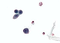
Photo from wikipedia
Background: Human polyomavirus (HPyV)6 and HPyV7 are shed chronically from human skin. HPyV7, but not HPyV6, has been linked to a pruritic skin eruption of immunosuppression. Objective: We determined whether… Click to show full abstract
Background: Human polyomavirus (HPyV)6 and HPyV7 are shed chronically from human skin. HPyV7, but not HPyV6, has been linked to a pruritic skin eruption of immunosuppression. Objective: We determined whether biopsy specimens showing a characteristic pattern of dyskeratosis and parakeratosis might be associated with polyomavirus infection. Methods: We screened biopsy specimens showing “peacock plumage” histology by polymerase chain reaction for HPyVs. Cases positive for HPyV6 or HPyV7 were then analyzed by immunohistochemistry, electron microscopy, immunofluorescence, quantitative polymerase chain reaction, and complete sequencing, including unbiased, next‐generation sequencing. Results: We identified 3 additional cases of HPyV6 or HPyV7 skin infections. Expression of T antigen and viral capsid was abundant in lesional skin. Dual immunofluorescence staining experiments confirmed that HPyV7 primarily infects keratinocytes. High viral loads in lesional skin compared with normal‐appearing skin and the identification of intact virions by both electron microscopy and next‐generation sequencing support a role for active viral infections in these skin diseases. Limitation: This was a small case series of archived materials. Conclusion: We have found that HPyV6 and HPyV7 are associated with rare, pruritic skin eruptions with a distinctive histologic pattern and describe this entity as “HPyV6‐ and HPyV7‐associated pruritic and dyskeratotic dermatoses.”
Journal Title: Journal of the American Academy of Dermatology
Year Published: 2017
Link to full text (if available)
Share on Social Media: Sign Up to like & get
recommendations!