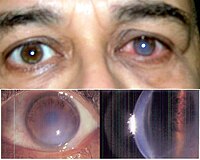
Photo from wikipedia
PURPOSE To investigate the efficacy of corneal crosslinking (CXL) for pellucid marginal degeneration (PMD). SETTING Medical University of Vienna. DESIGN Retrospective study. METHODS In eyes with a crab-claw pattern on… Click to show full abstract
PURPOSE To investigate the efficacy of corneal crosslinking (CXL) for pellucid marginal degeneration (PMD). SETTING Medical University of Vienna. DESIGN Retrospective study. METHODS In eyes with a crab-claw pattern on corneal topography of a study cohort of 808 eyes, manual measurements of the cornea's thinnest point on the inferior vertical Scheimpflug image (mCTi) and on the superior vertical Scheimpflug image (mCTs) were conducted. Eyes with paralimbal thinning were supposed as having PMD and included. A ratio between mCTi and mCTs was calculated. CXL was performed by irradiation of the inferior periphery of the cornea. During the follow-up, the mCTi, the mean keratometry (K) values in a central zone of 5.0 mm and in a 2.5 mm zone of the inferior cornea and the topographical corneal astigmatism were measured. The corrected distance visual acuity (CDVA) was also evaluated. Patients were followed postoperatively for 12 months. RESULTS Forty-eight eyes showed a crab-claw pattern in corneal topography. Twenty-two eyes matched the inclusion criteria for PMD and 16 eyes underwent CXL. The mCTi increased during the 12-month follow-up. The K value in the 2.5 mm zone of the inferior cornea decreased after 1 year, whereas the K value in the central zone of 5.0 mm remained stable. The corneal astigmatism continuously decreased, and the CDVA improved after 1 year. CONCLUSION Manual pachymetric measurements in Scheimpflug images showed the potential for screening for PMD and the evaluation of the efficacy of CXL in eyes with PMD in the study cohort. A thickening of the mCTi and a flattening in the inferior part of the cornea was observed.
Journal Title: Journal of cataract and refractive surgery
Year Published: 2019
Link to full text (if available)
Share on Social Media: Sign Up to like & get
recommendations!