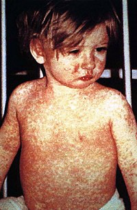
Photo from wikipedia
Fig 1. Case 1. Patch of alopecia on the occipital aspect of the scalp demonstrating a large, irregular zone of marked hair loss with intact follicular ostia and abrupt transition… Click to show full abstract
Fig 1. Case 1. Patch of alopecia on the occipital aspect of the scalp demonstrating a large, irregular zone of marked hair loss with intact follicular ostia and abrupt transition to normal scalp (image courtesy of Dr Arash Koochek). INTRODUCTION Alopecia areata typically presents with a distinctive histopathologic pattern, including peribulbar lymphocytic inflammation with or without occasional eosinophils, follicular miniaturization, and an increase in catagen/telogen hairs. Pigment incontinence, which is proportional to the pigmentation of the patient’s hair, is often present within the peribulbar fibrous root sheath. Only 3 cases of the granulomatous variant of alopecia areata have been reported, and we now describe 2 additional cases of granulomatous alopecia areata. This histologic variant of alopecia areata represents a potential pitfall when evaluating the histopathology of a patient with hair loss. Granulomatous inflammation should not preclude the diagnosis of alopecia areata in the appropriate clinical-pathologic setting.
Journal Title: JAAD Case Reports
Year Published: 2022
Link to full text (if available)
Share on Social Media: Sign Up to like & get
recommendations!