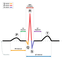
Photo from wikipedia
INTRODUCTION 3D mapping systems are used during radiofrequency (RF) pulmonary vein isolation (PVI) to facilitate catheter navigation and to provide additional electroanatomical information as a surrogate marker for the presence… Click to show full abstract
INTRODUCTION 3D mapping systems are used during radiofrequency (RF) pulmonary vein isolation (PVI) to facilitate catheter navigation and to provide additional electroanatomical information as a surrogate marker for the presence and location of fibrotic atrial myocardium. Electric voltage information can only be measured when the myocardium is depolarized. Low heart rates or frequent premature atrial beats can significantly prolong creation of detailed left atrial voltage maps. This study was designed to evaluate the potential advantage of voltage information collection during atrial pacing instead of acquisition during sinus rhythm. METHODS AND RESULTS A total of 40 patients were included in the study, in 20 consecutive patients voltage mapping was performed during sinus rhythm, and in the following 20 patients during atrial pacing. The average age of the included patients was 69.5 ± 9.4, 17 of 40 patients (43%) were male. All procedures were performed using the Carto 3D Mapping system. For LA voltage mapping, a multipolar circular mapping catheter was used. The atrium was paced via the proximal coronary sinus catheter electrodes with a fixed cycle length of 600 ms. By mapping during atrial pacing mapping time was reduced by 35% (441 s. (±141) vs. 683 s. (±203) p = 0.029) while a higher number of total mapping points were acquired (908 ± 560 vs. 581 ± 150, p = 0.008). CONCLUSION Acquiring left atrial low voltage maps during atrial pacing significantly reduces mapping time. As pacing also improves comparability of left atrial electroanatomical maps we suggest that this approach may be considered as a standard during these procedures.
Journal Title: Journal of electrocardiology
Year Published: 2020
Link to full text (if available)
Share on Social Media: Sign Up to like & get
recommendations!