Photo from wikipedia
BACKGROUND Little is known regarding the association between chronological out-stent vessel remodeling and in-stent tissue characteristics of drug-eluting stent (DES) failure. We aimed to evaluate the relationship between serial vessel… Click to show full abstract
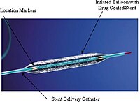
BACKGROUND Little is known regarding the association between chronological out-stent vessel remodeling and in-stent tissue characteristics of drug-eluting stent (DES) failure. We aimed to evaluate the relationship between serial vessel remodeling after DES implantation and neoatherosclerosis (NA) assessed by optical coherence tomography (OCT) in patients with DES failure. METHODS Forty-eight patients with late and very late stent failure after DES implantation, who underwent intravascular ultrasound (IVUS) at both the initial percutaneous coronary intervention and the time of stent failure and OCT imaging at the time of stent failure, were retrospectively investigated. NA on OCT was defined as neointimal formation with the presence of lipids or calcification inside the stents. Lesions were divided into two groups: those with NA and those without NA (NA: n=21; non-NA: n=27). From the serial IVUS examinations, external elastic membrane (EEM) volume and out-stent plaque volume were normalized by stent length and their changes were compared between the two groups. RESULTS The NA group showed older stent age [median, 5.1 years (IQR, 4.8-8.3) vs 1.4 years (IQR, 0.8-4.5); p<0.01] and more prevalent sirolimus-eluting stents (SES; 81.0% vs. 29.6%; p<0.01). IVUS findings of the NA group showed a greater serial increase in both normalized EEM volume and normalized out-stent plaque volume (OSPVI) [1.05 (0.41-1.90) vs. 0.11 (-0.64 to 0.80) mm2; p<0.01; and 0.88 (0.57-1.98) vs. 0.12 (-0.41 to 0.78) mm2; p<0.01]. On multivariate analysis, percentage change in OSPVI (OR, 1.07; 95% CI, 1.01-1.14; p=0.02) and SES (OR, 9.78; 95% CI, 2.20-43.40; p<0.01) remained independent predictors of NA. CONCLUSIONS NA in late and very late DES failure was associated with out-stent positive vessel remodeling. In addition to SES, out-stent progressive positive remodeling may help predict NA in late and very late DES failure.
Journal Title: Journal of cardiology
Year Published: 2018
Link to full text (if available)
Share on Social Media: Sign Up to like & get
recommendations!