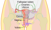
Photo from wikipedia
STUDY OBJECTIVE To evaluate the histology of the uterine septum after its complete hysteroscopic excision. DESIGN Case series. SETTING II Gynecological and Obstetric Unit of the University of Bari, Italy… Click to show full abstract
STUDY OBJECTIVE To evaluate the histology of the uterine septum after its complete hysteroscopic excision. DESIGN Case series. SETTING II Gynecological and Obstetric Unit of the University of Bari, Italy and Pathological Anatomy Department of the University of Bari, Italy. PATIENTS 35 patients between 25 and 41 years old diagnosed with uterine septum by 3D ultrasound as per ESHRE/ESGE 2013/Salim 2003 criteria. Office hysteroscopy was also performed to define the anatomy of the uterine cavity and to exclude the presence of other endometrial pathology. INTERVENTIONS Operative hysteroscopic septum resection was performed. The septum was initially incised with a "L shape" bipolar electrode with the 5 mm Bipolar Mini-Resectoscope (Karl Storz, Germany). Then, using the bipolar loop, two triangles of the septum were excised in parallel, obtaining uninterrupted entire septum-long strips, from the fundus to the apex of the septum. The strips were immediately removed from the uterus and reassembled "in vitro" in order to reconstruct the macroscopic 3D structure of the septum for a complete morphological and histological evaluation. MEASUREMENTS AND MAIN RESULTS Patients presented an average BMI of 24,8 and were all nulliparous. Histologic evaluation of the uterine septa showed a different conformation of the muscle bundles along the septum. In the apex and edges of the septum, the muscle cells were arranged in nodules circumscribed by a thin area of collagen fibers. Medium size vessels were distributed in the collagen fibers around the muscle cells. Only few capillary vessels are present in the muscle nodules. This pattern is very similar to the cell arrangement of leiomyomas. In the core of the septa, near the base, the muscle bundles show a linear course, with concurrent collagen fibers and vessels. All the above-mentioned characteristics were consistently present in every patient. On high-power histological fields (200x), the muscle portion ranged from 48,3 ± 1,8% (mean 6,0) in the apex and borders, to 48,5 ± 1,3% (mean 6,0) in the core. The collagen fibers ranged from 27.1 ± 1.1% (mean 4,0) in the apex and borders, to 26.7 ± 1.3% (mean 5,0) in the core. CONCLUSION This study allowed us, by removing the entire septum as a whole structure, to redefine the concept of the septum as a complex structure based on islands of muscle fibers irregularly arranged in vertex, in a context of collagen tissue, similar to the structure of myomas. It appears to deeply involve the anterior and posterior uterine wall, resembling a "reverse letter H".
Journal Title: Journal of minimally invasive gynecology
Year Published: 2019
Link to full text (if available)
Share on Social Media: Sign Up to like & get
recommendations!