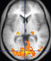
Photo from wikipedia
BACKGROUND The aims of this study were to assess the feasibility of magnetic resonance imaging (MRI) to track the in vivo distribution of autologous, injected blood in a subarachnoid hemorrhage… Click to show full abstract
BACKGROUND The aims of this study were to assess the feasibility of magnetic resonance imaging (MRI) to track the in vivo distribution of autologous, injected blood in a subarachnoid hemorrhage model (SAH), and to evaluate whether this technique results in observable morphological detriment. NEW METHOD We used an SAH model of stereotactic injection of autologous blood into the prechiasmatic cistern in Sprague Dawley rats. To visualize its in vivo distribution, a gadolinium-containing contrast agent was added to the autologous blood prior to injection. MRI was performed on a 9.4 T Bruker Biospec scanner preoperatively, as well as at variable time points between 30 minutes to 23 days after SAH. T1-weighted and diffusion-weighted images were acquired. The morphological examination was completed by a histopathological work-up. RESULTS Upon injection of contrast agent-enriched autologous blood, enhancement was observed in the entire subarachnoid space within 30 minutes of injection. Total clearance was noted at the first postoperative day. SAH induction did not result in changes in clinical scores or on histopathological or radiological images. COMPARISON WITH EXISTING METHODS We modified an established method to allow in vivo MRI monitoring of subarachnoid blood distribution in an SAH model. CONCLUSION This technique could be used to evaluate the distribution of blood components during the development of novel SAH models. Since no additional morphological detriment was observed, this technique could be used as a validation tool to verify correct application and induction in preclinical SAH models.
Journal Title: Journal of Neuroscience Methods
Year Published: 2019
Link to full text (if available)
Share on Social Media: Sign Up to like & get
recommendations!