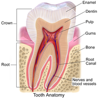
Photo from wikipedia
INTRODUCTION Idiopathic bone cavity (IBC) is an uncommon bone lesion that usually affects youngsters as an unilocular radiolucency with predilection for the posterior mandible. As the lesion is frequently located… Click to show full abstract
INTRODUCTION Idiopathic bone cavity (IBC) is an uncommon bone lesion that usually affects youngsters as an unilocular radiolucency with predilection for the posterior mandible. As the lesion is frequently located in proximity to the adjacent teeth, chronic apical periodontitis is commonly included as a differential diagnosis. The aim of the present study was to analyze the clinical and radiological features of a series of IBC diagnosed in a single service. METHODS All cases diagnosed as IBC were retrieved from the files of an Oral Pathology laboratory and the clinical and radiological characteristics were described with focus on differential diagnosis with chronic apical periodontitis. RESULTS Thirty cases composed the final sample; mean age of the affected patients was 22 years-old, there were no gender predilection and most lesions were located on the posterior (47%) and anterior (43%) mandible. Most lesions presented as unilocular radiolucencies (87%) and 90% were located in close association with the adjacent teeth. The associated teeth presented no endodontic involvement and all proved to be vital. CONCLUSIONS IBC usually affect young patients as an unilocular radiolucency in close association with the adjacent teeth. Careful radiological analysis and vitality tests of the adjacent teeth are essential to rule out chronic apical periodontis avoiding any unnecessary endodontic treatment.
Journal Title: Journal of endodontics
Year Published: 2020
Link to full text (if available)
Share on Social Media: Sign Up to like & get
recommendations!