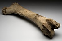
Photo from wikipedia
Summary Background/Objective The microstructure of the subchondral trabecular bone, including the composition and distribution of plates and rods, has an important influence on the disease progression and mechanical properties of… Click to show full abstract
Summary Background/Objective The microstructure of the subchondral trabecular bone, including the composition and distribution of plates and rods, has an important influence on the disease progression and mechanical properties of osteoarthritis (OA) and osteoporosis (OP). We aimed to determine whether differences in plates and rods influence the variations in the quantities and qualities of the subchondral trabecular bone between OA and OP. Materials and methods Thirty-eight femoral head samples [OA, n = 13; OP, n = 17; normal control (NC), n = 8] were collected from male patients undergoing total hip arthroplasty. They were scanned using microcomputed tomography, and subchondral trabecular structures were analysed using individual trabecular segmentation. Micro-finite element analysis (μFEA) was applied to assess the mechanical property of the trabecular bone. Cartilage changes were evaluated by using histological assessment. Analysis of variance was used to compare intergroup differences in structural and mechanical properties and cartilage degradation. Pearson analysis was used to evaluate the relationship between the trabecula microstructure and biomechanical properties. Results Compared with the OP and NC group, there was serious cartilage damage in the OA group. With respect to the microstructure results, the OA group had the highest plate and rod trabecular microstructures including number and junction density among the three groups. For the mechanical properties detected via μFEA, the OA group had higher stiffness and failure load than did the OP group. Pearson analysis revealed that compared with OP, OA had a higher number of microstructure parameters (e.g., rod bone volume fraction and rod trabecular number) that were positively correlated with its mechanical property. Conclusions Compared with OP, the OA subchondral bone has both increased plate and rod microarchitecture and has more microstructures positively related with its mechanical property. These differences may help explain the variation in mechanical properties between these bone diseases. The translational potential of this article Our findings suggested that changes in the plates and rods of the subchondral trabecular bone play a critical role in OA and OP progression and that the improvement of the subchondral trabecular bone may be a promising treatment approach.
Journal Title: Journal of Orthopaedic Translation
Year Published: 2020
Link to full text (if available)
Share on Social Media: Sign Up to like & get
recommendations!