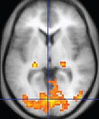
Photo from wikipedia
OBJECTIVE Over the last years several studies reported an increased signal intensity (SI) of the dentate nucleus (DN) on unenhanced T1-weighted images after repeated application of gadolinium-based contrast agents (GBCAs),… Click to show full abstract
OBJECTIVE Over the last years several studies reported an increased signal intensity (SI) of the dentate nucleus (DN) on unenhanced T1-weighted images after repeated application of gadolinium-based contrast agents (GBCAs), suggesting gadolinium deposition. The aim of this study was to investigate with diffusion-weighted MRI possible tissue abnormalities of the DN in multiple sclerosis (MS) patients. MATERIAL AND METHODS We retrospectively identified seventeen patients with at least six contrast-enhanced MRI examinations by using the linear GBCA gadopentate dimeglumine and twenty-three patients with the exclusive use of the macrocyclic contrast agent gadoterate meglumine followed by another 3 Tesla MRI scan including unenhanced T1-weighted and diffusion-weighted images. RESULTS In the linear GBCA group, we found significant differences of the DN-to-pons SI ratio on unenhanced T1-weighted images (1.13 ± 0.05) when compared to the macrocyclic GBCA group (0.97 ± 0.03; p < 0.001). However, we found no significant differences between apparent diffusion coefficient (ADC) values of the DN in both groups (linear GBCA group: 0.82 ± 0.04 × 10⁻³ mm/s2; marcocyclic GBCA group: 0.79 ± 0.04 × 10⁻³ mm/s2; p = 0.15). CONCLUSIONS Our results do not suggest that there is any difference in ADC values in the T1-hyperintense DN, which does not indicate a difference in tissue integrity between patients exposed to macrocyclic or linear GBCAs.
Journal Title: Magnetic resonance imaging
Year Published: 2019
Link to full text (if available)
Share on Social Media: Sign Up to like & get
recommendations!