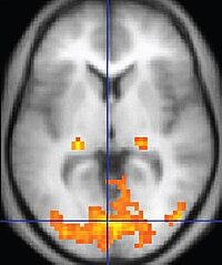
Photo from wikipedia
The thalamus serves as the central relay station for the brain. It processes and relays sensory and motor signals between different subcortical regions and the cerebral cortex and it can… Click to show full abstract
The thalamus serves as the central relay station for the brain. It processes and relays sensory and motor signals between different subcortical regions and the cerebral cortex and it can be divided into several neuronal clusters referred to as nuclei. Each of these can possibly be subdivided into sub-nuclei. Accurate and reliable identification of thalamic nuclei is important for surgical interventions and neuroanatomical studies. This is however a challenging task because the small size of the nuclei and the lack of contrast over the thalamus region in clinically acquired images does not permit the visualization of their boundaries. A number of methods have been developed for thalamus parcellation but the vast majority of these relies on diffusion imaging or functional imaging. The low resolution of these images only permit localizing the largest nuclei. In this work we propose a method to segment smaller nuclei. We first present a protocol to build histological-like atlases from a series of high-field (7 Tesla) MR images acquired with different pulse sequences that each permits to visualize the boundaries of a subset of the nuclei. We use this protocol to scan 9 subjects and we manually delineate 23 thalamic nuclei following the Morel atlas naming convention for each of these subjects. Manual contours for the nuclei are subsequently utilized to create statistical shape models. With these data, we compare four methods for the segmentation of thalamic nuclei in 3 T images we have also acquired for the 9 subjects included in the study: (1) single atlas, (2) multi atlas, (3) statistical shape, and (4) hierarchical statistical shape in which thalamic nuclei are hierarchically fitted to the images, starting from the largest ones. Results of a leave-one-out validation study conducted on the nine image sets we have acquired show that the multi atlas approach improves upon the single atlas approach for most nuclei. Segmentations obtained with the hierarchical statistical shape model yield the highest accuracy, with dice coefficients ranging from 0.53 to 0.90, mean surface errors from 0.27 mm to 0.64 mm, and maximum surface errors from 1.31 mm to 2.52 mm for all nuclei averaged across test cases. This suggests the feasibility of using such approach for localizing thalamic substructures in clinically acquired MR volumes. It may have a direct impact on surgeries such as Deep Brain Stimulation procedures that require the implantation of stimulating electrodes in specific thalamic nuclei.
Journal Title: Magnetic resonance imaging
Year Published: 2019
Link to full text (if available)
Share on Social Media: Sign Up to like & get
recommendations!