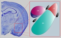
Photo from wikipedia
ABSTRACT The central nucleus of the amygdala (CeA) and bed nucleus of the stria terminalis (BNST), two nuclei within the central extended amygdala, function as critical relays within the distributed… Click to show full abstract
ABSTRACT The central nucleus of the amygdala (CeA) and bed nucleus of the stria terminalis (BNST), two nuclei within the central extended amygdala, function as critical relays within the distributed neural networks that coordinate sensory, emotional, and cognitive responses to threat. These structures have overlapping anatomical projections to downstream targets that initiate defensive responses. Despite these commonalities, researchers have also proposed a functional dissociation between the CeA and BNST, with the CeA promoting responses to discrete stimuli and the BNST promoting responses to diffuse threat. Intrinsic functional connectivity (iFC) provides a means to investigate the functional architecture of the brain, unbiased by task demands. Using ultra‐high field neuroimaging (7‐Tesla fMRI), which provides increased spatial resolution, this study compared the iFC networks of the CeA and BNST in 27 healthy individuals. Both structures were coupled with areas of the medial prefrontal cortex, hippocampus, thalamus, and periaqueductal gray matter. Compared to the BNST, the bilateral CeA was more strongly coupled with the insula and regions that support sensory processing, including thalamus and fusiform gyrus. In contrast, the bilateral BNST was more strongly coupled with regions involved in cognitive and motivational processes, including the dorsal paracingulate gyrus, posterior cingulate cortex, and striatum. Collectively, these findings suggest that responses to sensory stimulation are preferentially coordinated by the CeA and cognitive and motivational responses are preferentially coordinated by the BNST. HIGHLIGHTSThe CeA and BNST have overlapping functional connections.The CeA is more strongly connected to regions involved in sensory processing.The BNST is more strongly connected to regions involved in motivational processing.
Journal Title: NeuroImage
Year Published: 2018
Link to full text (if available)
Share on Social Media: Sign Up to like & get
recommendations!