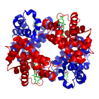
Photo from wikipedia
Highlights • Dopaminergic neurons dominate effective transverse relaxation in nigrosome 1.• Ion beam microscopy reveals highest iron concentrations in dopaminergic neurons.• Developed biophysical model links MRI parameters to cellular iron… Click to show full abstract
Highlights • Dopaminergic neurons dominate effective transverse relaxation in nigrosome 1.• Ion beam microscopy reveals highest iron concentrations in dopaminergic neurons.• Developed biophysical model links MRI parameters to cellular iron content.• Ferritin- and neuromelanin-bound iron impact MRI parameters differently.• Quantitative MRI provides a potential biomarker of iron in dopaminergic neurons.
Journal Title: Neuroimage
Year Published: 2021
Link to full text (if available)
Share on Social Media: Sign Up to like & get
recommendations!