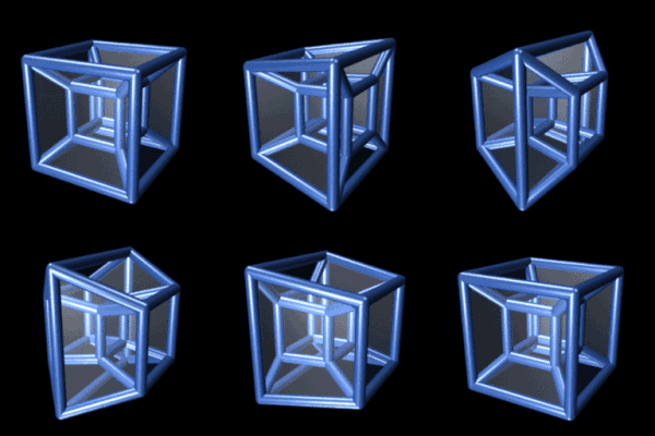
Photo from wikipedia
Abstract In order to overcome the shortcoming of traditional tomography requires the vast amount of data and the limitation of intersection angle between two beams existed in a microscope objective… Click to show full abstract
Abstract In order to overcome the shortcoming of traditional tomography requires the vast amount of data and the limitation of intersection angle between two beams existed in a microscope objective to realize the real-time detection of three-dimensional (3D) morphological distribution of blood cells in batches, a method of reconstructing cell substructure with only two non-orthogonal phases is proposed in this paper. In this work, an optimized maximum entropy tomography (MET) algorithm is used for rapid 3D reconstruction which requires less phase information from non-orthogonal directions. Moreover, two phase images can be obtained simultaneously by the phase imaging system combined with flow cytometry and optical tweezers (OT). We perform simulations of two types of cell models and experiments of red blood cell (RBC), thrombocyte and lymphocyte. Results demonstrate this method is of great significance for 3D morphological analysis of blood cells in the field of clinical diagnosis or even life sciences.
Journal Title: Optics and Lasers in Engineering
Year Published: 2020
Link to full text (if available)
Share on Social Media: Sign Up to like & get
recommendations!