Photo from wikipedia
OBJECTIVES VELscope® was developed to inspect oral mucosa autofluorescence. However, its accuracy is heavily dependent on the examining physician's experience. This study was aimed toward the development of a novel… Click to show full abstract
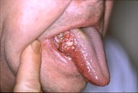
OBJECTIVES VELscope® was developed to inspect oral mucosa autofluorescence. However, its accuracy is heavily dependent on the examining physician's experience. This study was aimed toward the development of a novel quantitative analysis of autofluorescence images for oral cancer screening. MATERIALS AND METHODS Patients with either oral cancer or precancerous lesions and a control group with normal oral mucosa were enrolled in this study. White light images and VELscope® autofluorescence images of the lesions were taken with a digital camera. The lesion in the image was chosen as the region of interest (ROI). The average intensity and heterogeneity of the ROI were calculated. A quadratic discriminant analysis (QDA) was utilized to compute boundaries based on sensitivity and specificity. RESULTS 47 oral cancer lesions, 54 precancerous lesions, and 39 normal oral mucosae controls were analyzed. A boundary of specificity of 0.923 and a sensitivity of 0.979 between the oral cancer lesions and normal oral mucosae were validated. The oral cancer and precancerous lesions could also be differentiated from normal oral mucosae with a specificity of 0.923 and a sensitivity of 0.970. CONCLUSION The novel quantitative analysis of the intensity and heterogeneity of VELscope® autofluorescence images used in this study in combination with a QDA classifier can be used to differentiate oral cancer and precancerous lesions from normal oral mucosae.
Journal Title: Oral oncology
Year Published: 2017
Link to full text (if available)
Share on Social Media: Sign Up to like & get
recommendations!