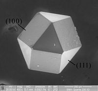
Photo from wikipedia
Abstract The microstructure of annealed single crystal 6H-SiC implanted with C+ and H2+ ions were characterized by glancing incidence X-ray diffraction (GIXRD), scanning electron microscopy (SEM) and transmission electron microscopy… Click to show full abstract
Abstract The microstructure of annealed single crystal 6H-SiC implanted with C+ and H2+ ions were characterized by glancing incidence X-ray diffraction (GIXRD), scanning electron microscopy (SEM) and transmission electron microscopy (TEM). After annealing, cracks were observed to form in the surface of C+ ions implanted sample. However, blisters as circular clusters were observed in the surface of the H2+ ions implanted and annealed sample. Compared with the samples implanted with only single type of ions, both cracks and blisters with different morphologies were found to appear in the surface of C+ and H2+ ions co-implanted sample. The morphology and distribution of blisters and exfoliations in the C+ and H2+ co-implanted samples were more irregular and inhomogeneous than those in the H2+ ions implanted sample, which can be attributed to the randomly distributed columnar crystals formed during the annealing process. The grain boundaries of these columnar crystals could be used as migration channels for hydrogen, which influence the size evolution of blisters and exfoliations in the sample surface during annealing process.
Journal Title: Progress in Nuclear Energy
Year Published: 2020
Link to full text (if available)
Share on Social Media: Sign Up to like & get
recommendations!