Photo from wikipedia
BACKGROUND Traumatic brain injury (TBI) lesions are known to evolve over time, but the duration and consequences of cerebral remodeling are unclear. Degenerative mechanisms occurring in the chronic phase after… Click to show full abstract
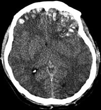
BACKGROUND Traumatic brain injury (TBI) lesions are known to evolve over time, but the duration and consequences of cerebral remodeling are unclear. Degenerative mechanisms occurring in the chronic phase after TBI could constitute "tertiary" lesions related to the neurological outcome. OBJECTIVE The objective of this prospective study of severe TBI was to longitudinally evaluate the volume of white and grey matter structures and white matter integrity with 2 time-point multimodal MRI. METHODS Longitudinal MRI follow-up was obtained for 11 healthy controls (HCs) and 22 individuals with TBI (mean [SD] 60 [15] months after injury) along with neuropsychological assessments. TBI individuals were classified in the "favourable" recovery group (Glasgow Outcome Scale Extended [GOSE] 6-8) and "unfavourable" recovery group (GOSE 3-5) at 5 years. Variation in brain volumes (3D T1-weighted image) and white matter integrity (diffusion tensor imaging [DTI]) were quantitatively assessed over time and used to predict neurological outcome. RESULTS TBI individuals showed a marked decrease in volumes of whole white matter (median -11.4% [interquartile range -5.8; -14.6]; p <0.001) and deep grey nuclear structures (-17.1% [-10.6; -20.5]; p <0.001). HCs did not show any significant change over the same time period. Median volumetric loss in several brain regions was higher with GOSE 3-5 than 6-8. These lesions were associated with lower fractional anisotropy and higher mean diffusivity at baseline. Volumetric variations were positively correlated with normalized fractional anisotropy and negatively with normalized mean diffusivity at baseline and follow-up. A computed predictive model with baseline DTI showed good accuracy to predict neurological outcome (area under the receiver operating characteristic curve 0.82 [95% confidence interval 0.81-0.83]) Conclusions. We characterised the striking atrophy of deep brain structures after severe TBI. DTI imaging in the subacute phase can predict the occurrence and localization of these tertiary lesions as well as long-term neurological outcome. TRIAL REGISTRATION ClinicalTrials.gov: NCT00577954. Registered on October 2006.
Journal Title: Annals of physical and rehabilitation medicine
Year Published: 2021
Link to full text (if available)
Share on Social Media: Sign Up to like & get
recommendations!