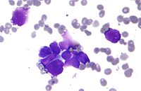
Photo from wikipedia
We report a case of a huge solitary non-endobronchial pulmonary tumor in a 76-year-old male smoker. The tumor measured 11 × 10 × 8 cm. It was ill-defined, and it was located periphery of… Click to show full abstract
We report a case of a huge solitary non-endobronchial pulmonary tumor in a 76-year-old male smoker. The tumor measured 11 × 10 × 8 cm. It was ill-defined, and it was located periphery of the right lower lobe with the subpleural cystic spaces. He underwent right lower lobectomy with mediastinal lymph node dissection and is free from tumor 30 months after surgery. Microscopically, it was composed of a proliferation of squamous and ciliated columnar epithelial cells with a few mucous cells. These cells were arranged in a papillary growth fashion extending along the fibrously thickened alveolar septa together with metaplastic bronchiolar and squamous epithelia displaying an usual interstitial pneumonia-pattern. Although the histologic features of the tumor were that of a mixed squamous cell and glandular papilloma (MSCGP), it was peripherally located and showed a lepidic growth, and it was much larger than previously reported MSCGPs. It is possible that the tumor developed in association with bronchial metaplasia in the periphery of the lung, and then extended along the surface of the reconstructed air spaces, which resulted in its unique histologic appearance. Further investigations of respiratory papilloma are needed to clarify the pathogenesis of these lesions.
Journal Title: Respiratory Medicine Case Reports
Year Published: 2018
Link to full text (if available)
Share on Social Media: Sign Up to like & get
recommendations!