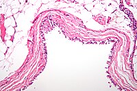
Photo from wikipedia
BACKGROUND Small, incidental filar cyst associated with terminal lipoma is thought to be caused by failure of secondary neurulation; however, the precise embryological background is not fully understood. Retained medullary… Click to show full abstract
BACKGROUND Small, incidental filar cyst associated with terminal lipoma is thought to be caused by failure of secondary neurulation; however, the precise embryological background is not fully understood. Retained medullary cord (RMC) also originates from late arrest of secondary neurulation. The central feature of RMC histopathology is a central canal-like ependyma-lined lumen with surrounding neuroglial core. CASE DESCRIPTION We surgically treated two patients with a large cyst in the rostral part of the filum and lipoma in the caudal filum. At cord untethering surgery, the filum was severed at the caudal part of the cyst. Histopathologically, the filar cyst was the cystic dilatation of the central canal-like structure at the marginal part of the lipoma. The central canal-like structure was continuous caudally in the lipoma and its size decreased towards the caudal side. CONCLUSION The present findings support the idea raised by Pang et al. that entities such as filar cyst, terminal lipomas, and RMC can all be considered consequences of a continuum of regression failure occurring during late secondary neurulation.
Journal Title: World neurosurgery
Year Published: 2020
Link to full text (if available)
Share on Social Media: Sign Up to like & get
recommendations!