Photo from wikipedia
Objective Improved understanding of the time course of neural changes associated with adolescent PTSD would elucidate the development of the disorder and could inform approaches to treatment. We compared hippocampal… Click to show full abstract
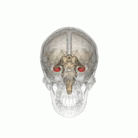
Objective Improved understanding of the time course of neural changes associated with adolescent PTSD would elucidate the development of the disorder and could inform approaches to treatment. We compared hippocampal volumes and resting state functional connectivity (RSFC) in adolescent girls with post-traumatic stress disorder (PTSD) secondary to sexual assault, within six months of onset and age- and gender-matched, non-trauma exposed healthy controls (HCs) in São Paulo, Brazil. We also examined the relationship between pre- and post-treatment PTSD symptoms and RSFC. Method We collected brain structure, RSFC, and PTSD symptoms in 30 adolescents with PTSD (mean age: 15.7 ± 1.04 years) and 21 HCs (mean age: 16.2 ± 1.21 years) at baseline. We collected repeated measures in 21 participants with PTSD following treatment; 9 participants dropped out. Hippocampal volume and RSFC from hippocampal and default mode network (DMN) seeds were compared between participants with PTSD and HCs. We examined associations between within-subject changes in RSFC and PTSD symptoms following treatment. Results No hippocampal volumetric differences between groups were found. Compared to HCs, adolescents with recent PTSD had reduced RSFC between hippocampus and the lateral parietal node of the DMN, encompassing the angular gyrus, peak coordinates: −38, −54, 16; 116 voxels; peak F1,47 = 31.76; FDR corrected p = 0.038. Improvements in PTSD symptoms were associated with increased RSFC between hippocampus and part of the lateral parietal node of the DMN, peak coordinates: −38, −84, 38; 316 voxels; peak F1,47 = 40.28; FDR corrected p < 0.001. Conclusion Adolescents with recent PTSD had reduced hippocampal-DMN RSFC, while no group differences in hippocampal volume were found, suggesting that hippocampal function, but not structure, is altered early in the course of PSTD. Following treatment, hippocampal-DMN RSFC increased with symptom improvement and may indicate an important neural mechanism related to successful PTSD treatment.
Journal Title: Neurobiology of Stress
Year Published: 2022
Link to full text (if available)
Share on Social Media: Sign Up to like & get
recommendations!