Photo from wikipedia
Abstract Background Larger grey matter volume of the inferior frontal gyrus (IFG) is among the most replicated biomarkers of genetic risk for bipolar disorders (BD). However, the IFG is a… Click to show full abstract
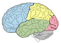
Abstract Background Larger grey matter volume of the inferior frontal gyrus (IFG) is among the most replicated biomarkers of genetic risk for bipolar disorders (BD). However, the IFG is a heterogeneous prefrontal region, and volumetric findings can be attributable to changes in cortical thickness (CT), surface area (SA) or gyrification. Here, we investigated the morphometry of IFG in participants at genetic risk for BD. Methods We quantified the IFG cortical grey matter volume in 29 affected, 32 unaffected relatives of BD probands, and 42 controls. We then examined SA, CT, and cortical folding in subregions of the IFG. Results We found volumetric group differences in the right IFG, with the largest volumes in unaffected high-risk and smallest in control participants (F2,192 = 3.07, p = 0.01). The volume alterations were localized to the pars triangularis of the IFG (F2,97 = 4.05, p = 0.02), with no differences in pars opercularis or pars orbitalis. Pars triangularis volume was highly correlated with its SA [Pearson r(101) = 0.88, p < 0.001], which significantly differed between the groups (F2,97 = 4.45, p = 0.01). As with volume, the mean SA of the pars triangularis was greater in unaffected (corrected p = 0.02) and affected relatives (corrected p = 0.05) compared with controls. We did not find group differences in pars triangularis CT or gyrification. Conclusions These findings strengthen prior knowledge about the volumetric findings in this region and provide a new insight into the localization and topology of IFG alterations. The unique nature of rIFG morphology in BD, with larger volume and SA early in the course of illness, could have practical implications for detection of participants at risk for BD.
Journal Title: Psychological Medicine
Year Published: 2018
Link to full text (if available)
Share on Social Media: Sign Up to like & get
recommendations!