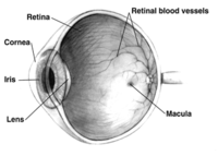
Photo from wikipedia
The axons of retinal ganglion cells (RGCs) comprise the optic nerve (ON)[1]. The postnatal ON in humans and rodents grows in length by over 80% [2]. However, the mechanism of… Click to show full abstract
The axons of retinal ganglion cells (RGCs) comprise the optic nerve (ON)[1]. The postnatal ON in humans and rodents grows in length by over 80% [2]. However, the mechanism of postnatal axonal growth is still poorly understood, and has been hypothesized by some to occur diffusely, since axon myelination has already occurred. We recently determined that the most anterior portion of the ON in mammals (the optic nerve lamina region, ONLR) contains a SOX2(+) neural progenitor cell (NPC) niche that can ultimately give rise to oligodendrocyte progenitors (OPCs), and all major glial forms: Type 1 and 2 astrocytes and oligodendrocytes. We wanted to determine the role of this SOX2(+)ONLR niche during the period of postnatal axonal growth. We used animals whose SOX2(+) cells would self-delete using a genetic construct that expresses Diphtheria toxin-Subunit A (DTA). Even a single molecule of DTA results in cell death within three days.
Journal Title: Microscopy and Microanalysis
Year Published: 2018
Link to full text (if available)
Share on Social Media: Sign Up to like & get
recommendations!