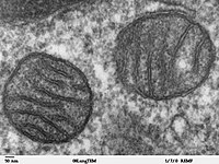
Photo from wikipedia
Several vital physiological processes, such as phospholipid and Ca2+ exchange, mitochondrial biogenesis, autophagy, and intracellular trafficking rely on the contact sites between the endoplasmic reticulum and mitochondria (Kornmann et al.,… Click to show full abstract
Several vital physiological processes, such as phospholipid and Ca2+ exchange, mitochondrial biogenesis, autophagy, and intracellular trafficking rely on the contact sites between the endoplasmic reticulum and mitochondria (Kornmann et al., 2009; Hirabayashi et al., 2017). These processes are affected in neurodegenerative disorders such as Alzheimer's (Area-Gomez and Schon, 2016), Parkinson's, amyotrophic lateral sclerosis (ALS), and frontotemporal dementia (FTD) (Paillusson et al., 2016). However, it is not clear how these disorders can affect multiple seemingly unrelated physiological pathways. ER-mitochondria contact sites are emerging as candidates in understanding the origins of the pathophysiology of these disorders. The contact sites are small sporadic areas where the two organelles are less than 30 nanometers apart. There are specialized protein complexes that reside at these sites. This results in a complex molecular landscape that is not fully understood. Several protein complexes have been proposed as the tethers that bridge these membranes. Tethers are comprised of ER-resident proteins, as well as mitochondrial outer membrane proteins, and in some cases soluble intermediate proteins. Here, the molecular composition and architecture of one of these tethers is studied in eukaryotic cells with endogenous fluorescence tags. One ER-resident protein, PDZD8, and one interacting partner, a mitochondrial outer membrane protein, are studied in vivo. Further, the structure of these macromolecular complexes is explored using cryo-EM. With state-of-the-art cryo-correlative light and electron microscopy, the distribution of the tethers as well as their structural mechanisms of mediating contact are uncovered and characterized.
Journal Title: Microscopy and Microanalysis
Year Published: 2021
Link to full text (if available)
Share on Social Media: Sign Up to like & get
recommendations!